FAQ on Apoptosis
The FAQ on Apoptosis:
In this blog, I will discuss about Definition of apoptosis, causes, morphological changes, biochemical changes, pathways, mechanisms, the difference between apoptosis and necrosis, etc.
Q 1: Definition of Apoptosis:
Ans: Apoptosis is a pathway of cell death that is induced by a tightly regulated suicide program in which cells destined to die to activate intrinsic enzymes that degrade the cells' own DNA and nuclear and cytoplasmic proteins. (Ref: Robbin's).
In a simplified way, Apoptosis is a way of programmed cell death in which specific enzymes are activated and there is the destruction of unwanted cells.
 |
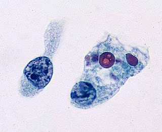
| Apoptosis |
Q 2: What are apoptotic bodies?
Ans: Apoptotic cells breaks up into cell fragments containing part of cytoplasm and nucleus of cell.These fragments are known as APOPTOTIC BODIES.
Q 3: What are the causses of Apoptosis?
Ans:
Apoptosis occurs in following physiologic conditions-
- Desruction of cells during embryogenesis.
- Regression of hormone-dependent tissues upon hormone with-drawl,such as- atresia of ovarian follicle in menopause,regressing of lactating breast tissue after stopage of breast feeding.
- Loss of cells in proliferating cell population- death of immature lymphocytes that fails toproduce antibodies, death of old epitheliial cells in intestinal crypts.
- Death of potentially harmful self-reactive lymphocytes.
- Death of neutrophils after acute inflammatory respone and lymphocytes after immune response.
Apoptosis also occurs in following pathological conditions:
- DNA damage: If radiation/cytotoxic drugs/hypoxia causes irreversible DNA damage,then there is activation of intrinsic mechanisms leadng to Apoptosis.
- Accumulation of improperly folded proteins in endoplasmic reticulum leads to a condition named ER stress,which also leads to APOPTOSIS.
- Apoptosis also occur in virus infected cells to control infection.
- Pathologic atrophy of organ after blockage of duct in pancreas/parotid.
Q 4: What are the morphogical changes that are observed in apoptosis?
Ans:
a) Cell shrinkage: There is inncreased density of cytopllasm with tigtly packed organelles.
b) Chromatin condensation followed by nuclear fragmentation.
c) There is formation of cytoplasmic blebs followd by formation of membrane bound apoptoti bodies.
d) Finally there is phagocytosis of apoptooti bodies followed by lysosoomal degradation.
Q 5 : Sequence of biohemical events in apoptosis.
 |
| Biochemical events in Apoptosis |
Q 6: Mechanism of apoptosis.
Ans: Apoptosis results from activation of the enzyme Caspases. The process of apoptosis is divided into initiation phase and execution phase.The process of apoptosis is regulated by pro-apoptotic and anti-apoptotic proteins. Caspase activation can occur by mitochondrial pathway or death receptor pathway.
Mitochondrial or intrinsic pathway of apoptosis:
Inactive caspases resides in the cytoplasm and it is activated by CytochromeC, which actually resides in mitochondrial inter membrane space. Release of Cytochrome C into cytosol is tightly regulated by 3 groups of proteins:1) Anti-apoptotic 2) Pro-apoptotic 3) Sensors.
 |
| Intrinsic pathway of caspase activation |
On reaching the cytoplasm cytochrome C binds with a protein named APAF(Apoptosis activating factor) and forms a hexamer called apoptosome,that cleaves caspase9.Also there is release of Smac or Diablo from inter-mitochondrial membrane to cytosol that binds and inactivate inhibitors of apoptosis(IAPs)proteins.
The extrinsic or Death receptor initiated pathway of apoptosis:
This pathway is initiated by death receptor(a member of TNF receptor family).When there is interaction of Fas ligand with the Fas portion of receptor, the cytoplasmic portions of Fas receptors comes closer to form binding site for FADD and this interm leads to activation of caspase8(caspase10 in human).Finally all these leads to initiation of execution phase.
The execution phase of apoptosis:
The execution pathway is the final common pathway of apoptosis.At the start of execution pathway there is activation of caspase3.Due to activation of caspase3 endonuclease and protease enzymes are activated.Endonuclease causes degradation and fragmentation of nuclear DNA.Activation of protease causes degradation of cytoskeletal and nuclear proteins.So, there is disorganization of cellular structure leading to formation of apoptotic bodies.Finally these apoptotic bodies will be taken up by macrophages.
Q7: How apoptosis is diagnosed?
Ans: Apoptosis can be detected by the following methods:
- Electron microscopy.
- Proteomic and genomic analysis.
- Spectroscopic techniques.
- Flowcytometry.
- Caspase activity assay.
- Microfluidic application.
Q8: Difference between Apoptosis and Necrosis.
Ans:
 |
| Necrosis vs Apoptosis |
Most importantly, apoptosis involve single or a few cells at a time and there is no spillage of the cellular contents into the environment, so there is abscence of local inflammatory cell infiltration.But, in case of necrosis, there is involvement of group of cells along with spillage of cellular contents into the surrounding environment provoking local inflammatory cell infiltration.
 |
| Figure:Local inflammatory infiltrate in necrosis |
Q 9: What is Necroptosis?
Ans: We all know that necrosis is a form of unprogrammed cell death leading to outpouring of cellular content into extracellular environment and resulting in local inflammatory cell infiltration.But necroptosis is a form of programmed necrosis.
It is commonly observed during viral infection. Apoptosis is the usual pathway by which virus infected cells are destroyed to controll the infection.But sometimes some essential factors of apoptosis are blocked by viral proteins.In this scenario necroptosis occurs as a backup method for control of viral infection.TNF Alfa,TRADD are the key players in necroptosis.
CMV commonly express caspase inhibitors to prevent apoptosis. Recently necroptosis based therapies are emerging to fight cancer and some other diseases.
If you find this content helpful, then don't forget to share it in social media with others!!!!
Q 10: Role of apoptosis in cancer.
 |
| Apoptosis in cancer |
Ans: When the DNA of a cell is damaged due to some injurious agent, there maybe a chance of faulty repair. Some mutations can also occur. And this mutations can lead to cancer. So the cells with mutations are destined to die to prevent carcinogenesis.One of the way of this type of cell death is apoptosis. So decreased apoptosis can lead to cancer. There are few ways that were proved to cause reduced apoptosis and thereby causing cancer:
1) Disturbed balence between proapoptotic and antiapoptotic proteins:
BCL2 is an antiapoptotic protein which resides on the outer membrane of mitochondria and prevent apoptosis. In case if B cell lymphoma with translocation(14;18), there is overexpression of BCL 2 gene. So there is excessive production of BCL2 protein. Due to overproduction of this antiapoptotic protein, there is blockade of apoptosis and cancerous cells fails to die and finally results in lymphoma.Also due to mutation of p53 gene, there is decrease in apoptosis leading to developement of various cancers.
2) Overexpression of inhibitor of apoptosis proteins(IAPs):
There are some inhibitors of apoptosis such as survivin that are present in adequate amount in cytosol to balence cell death. But in some hematological malignancy or pancreatic cancer these proteins are overexpressed.
3) Reduction in caspase activity:
Caspases are essential enzymes in intrinsic, extrinsic as well as in execution pathway of apoptosis.Reduced caspase activity was observed in various breast, cervical and ovarian carcinomas.
4)Improper death receptor signalling:
There are various death receptors involved in extrinsic pathway of apoptosis like TNFR1, DR3, DR4 etc. In few leukaemia or neuroblastoma, there is downregulation of these death receptors.

Visit our website homepage for more articles on Pathology👇
www.pathologydiscussion.com
Feel free to share any of our articles on social media(Directly from the website) if you find it helpful. Thank you!!!!

Comments
Post a Comment
Thank you for posting your comment.Your question will be answered soon.