FAQ about NECROSIS: Definition, morphological changes,types like coagulative,liquefactive,Caseous,fat and fibrinoid necrosis
FAQ about NECROSIS: Detailed discussion about the definition, morphological changes different types such as coagulative, liquefactive,Caseous, fat and fibrinoid necrosis for USMLE, PLAB, NEET PG and other medical examinations.
Q 1. What is the definition of Necrosis?
Ans: "Necrosis can be defined as circumscribed death of cells or tissue with structural evidence of death occurring in a living body"
In a simplified way, when there is a disturbance in the surrounding environment beyond homeostasis, there is premature cell death-which is called Necrosis. Due to damage to the cell membrane there is leakage of the cellular contents into the extracellular spaces that elicit inflammation.
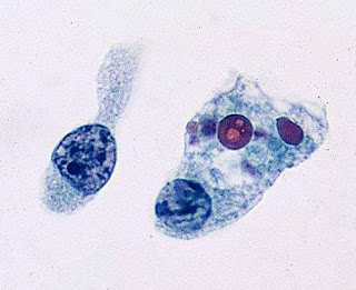
 |
| Necrosis: Spillage of intracellular contents |
Q 2. What are the morphological changes that occur in Necrosis?
Ans: Cytoplasmic changes:
- There is cytoplasmic eosinophilia due to the loss of cytoplasmic RNA denatured cytoplasmic proteins.

Cytoplasmic eosinophilia - The glassy homogenous appearance of the cells due to loss of glycogen particle.
- Whorled phospholipid masses are known as myelin figures derived from the damaged cell membrane.
- Marked dilation of mitochondria, aggregates of fluffy material and amorphous debris also noted.

- Karyolysis: There is the degradation of the DNA material by the endonuclease enzyme. So, there is less intensity of the blue color of the nucleus.
- Pyknosis: Due to the condensation of chromatin, there is increased basophilia of chromatin.
- Karyorrhexis: There is the fragmentation of the pyknotic nucleus.
Ans: It is a type of necrosis where the architecture of the dead tissue is preserved for a few days.
Here due to hypoxic insult, there is denaturation of the structural proteins as well as the enzymatic proteins, so there is no proteolysis. Morphologically cells appear as anucleate cells with pink cytoplasm. Coagulative necrosis can occur in any organ except the brain.
Q 4. What is liquefactive necrosis?
Ans: In this type of necrosis there is digestion of the dead cells leading to the formation of a liquid mass. It occurs in focal bacterial and occasionally in fungal infection. Due to these infections, there is an infiltration of leucocytes. From these leucocytes, there is the liberation of lysosomal enzymes and these enzymes are responsible for the autolysis of cells. Liquefactive necrosis is also known as "Wet gangrene".
Q 5. What is Fat necrosis?
Ans: It is the focal areas of fat destruction resulting from the release of pancreatic lipase into the peritoneal cavity. By action of pancreatic lipase, fatty acids are produced. Fatty acids combine with calcium to produce chalky white deposits.
Q 6. What is Fibrinoid necrosis?
Ans: This type of necrosis occurs when the immune complex(Antigen-Antibody complex) deposited along the wall of arteries. On microscopy, these deposits appear as fibrin lie bright pink deposits.
 |
| Fibrinoid necrosis |

Feel free to share any of our articles on social media(Directly from the website) if you find it helpful. Thank you!!

Comments
Post a Comment
Thank you for posting your comment.Your question will be answered soon.