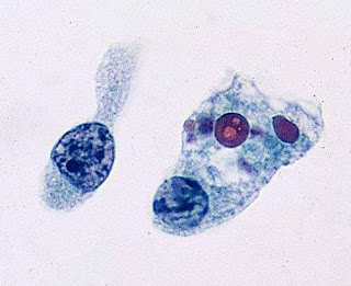
Found in a Papillary carcinoma thyroid slide!!!!
Yesterday while reviewing the slides of a Papillary carcinoma thyroid these cells were found adjacent to normal thyroid tissue.
Initially I thought ....what are these cells??
So...it is the best time to have a look at the histology of Parathyroid gland.
Histology of Parathyroid gland:
- Composed primarily of chief cells and fat with thin fibrous capsule dividing gland into lobules
- May have a pseudofollicle pattern resembling thyroid follicles (pink material is PAS positive)
- 6 - 8 microns, polygonal, central round nuclei, contain granules of parathyroid hormone (PTH)
- Basic cell type, other cell types are due to differences in physiologic activity
- 80% of chief cells have intracellular fat
- Chief cell is most sensitive to changes in ionized calcium
- Slightly larger than chief cell (12 microns), acidophilic cytoplasm due to mitochondria
- No secretory granules
- First appear at puberty as single cells, then pairs, then nodules at age 40
- Abundant optically clear cytoplasm and sharply defined cell membranes
- Chief cells with excessive cytoplasmic glycogen.
Okk...thats it for now....If you like this information, then dont forget to share it with others!!! and....
If you like, you can visit our youtube channel and facebook page👇🏻
Youtube channel link-https://m.youtube.com/channel/UCPGvHc5Ttw4EtB72WVMiYSw/videos
Facebook page link-https://m.facebook.com/PathologyDiscussionForum/?ref=bookmarks
If you like, you can visit our youtube channel and facebook page👇🏻
Youtube channel link-https://m.youtube.com/channel/UCPGvHc5Ttw4EtB72WVMiYSw/videos
Facebook page link-https://m.facebook.com/PathologyDiscussionForum/?ref=bookmarks
Follow our facebook page for recent articles/videos.🙂🙏🏻


Comments
Post a Comment
Thank you for posting your comment.Your question will be answered soon.