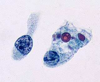
"Two-tone” cytoplasmic staining
Astonishing “two-tone” cytoplasmic staining pattern:
Radiation effect in cervicovaginal PAP smear. Radiation looks like a wild reparative reaction, with large cells, multinucleation, cytoplasmic vacuolization, and a curious “two-tone” cytoplasmic staining pattern(arrow)...........................
The characteristic changes are marked cellular and nuclear enlargement with preservation of the nuclearto- cytoplasmic ratio, cytoplasmic vacuolization, and cytoplasmic polychromasia (“two-tone” cytoplasm) . Nuclei have finely granular chromatin or show smudgy hyperchromasia, and there can be nuclear and cytoplasmic vacuolization. Cells are isolated or arranged in groups, and multinucleation is common. Reparative epithelium commonly accompanies radiation changes. Some chemotherapeutic drugs induce similar changes.Radiation changes superficially resemble herpes cytopathic changes. Multinucleation occurs in both
conditions, but radiation lacks the ground glass nuclear appearance or Cowdry A type inclusions typical of herpes.
If the radiation was given for a cervical cancer, the differential diagnosis includes recurrent SQC or adenocarcinoma of the cervix, with superimposed radiation changes. The cells of a recurrent SQC and adenocarcinoma
are typically more numerous than the scattered radiation cells. Recurrent cancers show more significant nuclear atypia than is seen in radiation. Coarsely textured chromatin (rather than smudgy hyperchromasia)
is typical of nonkeratinizing SQC.
(Ref:Cytology : diagnostic principles and clinical correlates / Edmund S. Cibas, Barbara S. Ducatman. — 3rd ed.)
If you like, you can visit our youtube channel and facebook page👇🏻
Youtube channel link-https://m.youtube.com/channel/UCPGvHc5Ttw4EtB72WVMiYSw/videos
Facebook page link-https://m.facebook.com/PathologyDiscussionForum/?ref=bookmarks
Follow our facebook page for recent articles/videos.


Comments
Post a Comment
Thank you for posting your comment.Your question will be answered soon.