
Hydatid Cyst:
In North America, hydatid disease is caused by ova of the tapeworm Echinococcus granulosus, a parasite of dogs and wolves. The ova are passed free in a dog’s or wolf ’s feces and develop into six-hooked embryos in the duodenum when swallowed by a human host. The embryos enter the venules and are filtered
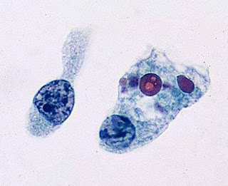
by the liver, developing into hydatid cysts that bear numerous scoleces provided with hooklets(green arrow), which represent the future heads of adult tapeworms.
Cytology:
The fluid aspirated from a hydatid cyst is usually clear and contains debris, a few inflammatory cells, and numerous scoleces. In old cysts, the aspirate yields large fragments of laminated layer of the cyst wall in a dirty background with debris containing detached hooklets and calcareous corpuscles. The scoleces may be difficult to find in FNA smears, but the hooklets in a ring-like arrangement or scattered in the debris often remain. The finding of hooklets or laminated layer of the cyst wall is diagnostic of hydatid disease.
Key features of hydatid liver cyst:
• CT scan: cystic mass.
• Dirty background containing fragments of laminated wall,
debris embedded with hooklets, and calcareous bodies at
low power.
• Scolex with hooklets seen at high power.

Comments
Post a Comment
Thank you for posting your comment.Your question will be answered soon.