Carcinoid
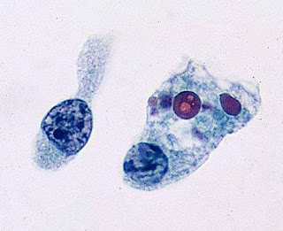
Beautiful picture showing Carcinoid tumor of the ileum.
 The tumor is composed of nests, glandular structures, and trabeculae of cells enmeshed in a fi brotic stroma. The cells are polarized around central lumina and have abundant, faintly eosinophilic cytoplasm and bland nuclei with stippled chromatin and small nucleoli.
Carcinoid tumors may have a variable gross appearance, ranging from the presence of polypoid tanyellow submucosal nodules to annular masses or strictures secondary to the marked fibrosis characteristic of these lesions. In approximately 20% of cases, the lesions may present as multiple mucosa-based polypoid lesions.153,160 X-chromosome inactivation analysis of multifocal carcinoid tumors of the small intestine has demonstrated identical X-chromosome inactivation patterns, indicating that multiple tumors represent mucosal metastases. Ileal carcinoid tumors are composed of nests and trabeculae of monotonous epithelial cells arranged in an organoid fashion and enmeshed within a fi brotic stroma.Th e tumor cells are cytologically bland with abundant amphophilic or faintly eosinophilic cytoplasm, round nuclei with stippled chromatin, and inconspicuous nucleoli. Necrosis, mitotic activity, and severe cytologic atypia are not characteristic of these tumors, although most show perineural and lymphovascular
invasion.(Ref: Odze surgical Pathology)
Visit Pathology discussion forum for more topics.

Comments
Post a Comment
Thank you for posting your comment.Your question will be answered soon.