Lipofuscin vs Cholestasis
Lipofuscin vs Cholestasis:

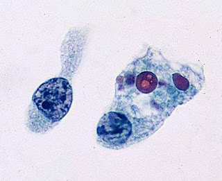
Comparison of lipofuscin pigment (left) and cholestasis (right), both found predominantly in zone 3. Lipofuscin pigment is fi ne, well delineated, light brown, and located particularly at the canalicular pole of hepatocytes. Intracellular bile in cholestasis has a greenish hue, is less granular, and often forms canalicular thrombi (arrows).
Lipofuscin pigment is normally seen in variable quantities in the centrilobular areas as periodic acid–Schiff (PAS)- positive, diastase-resistant, fi ne, light brown granules. There is a progressive increase of lipofuscin in individual hepatocytes with age.The granules represent lysosomes that contain materials that cannot be further degraded, and thus they are not present in recently regenerated hepatocytes. Intracellular bile, in cholestasis, can be distinguished from lipofuscin by its greenish hue, lack of a granular appearance, and its frequent formation of thrombi in bile canaliculi in zone 3 hepatocytes. Large amounts of lipofuscin are difficult to distinguish from Dubin-Johnson pigment microscopically. In comparison to lipofuscin, iron and copper are coarser, birefringent, and usually deposited in periportal hepatocytes. Hemosiderin and copper are abundant in the cytoplasm of hepatocytes during the fi rst week of life, then gradually disappear, and should be absent before the age of 9 months. Small quantities of stainable iron are common in normal hepatocytes,particularly in older individuals.(Ref:Odze Surgical Pathology)
If you like, you can visit our youtube channel and facebook page👇🏻
Youtube channel link-https://m.youtube.com/channel/UCPGvHc5Ttw4EtB72WVMiYSw/videos
Facebook page link-https://m.facebook.com/PathologyDiscussionForum/?ref=bookmarks
Follow our facebook page for recent articles/videos 😎

Comments
Post a Comment
Thank you for posting your comment.Your question will be answered soon.